Jose Espejo Valle-Inclán, formerly at the European Molecular Biology Laboratory’s European Bioinformatics Institute (EMBL-EBI) in Hinxton, United Kingdom, is now a Würth Research Centre of Hope for Pancreatic Cancer Group Leader based at the Botton-Champalimaud Pancreatic Cancer Centre at the Champalimaud Foundation. Vincenzo Corbo is an Associate Professor at the Department of Engineering for Innovation Medicine and ARC-Net Research Centre at the University of Verona, Italy, and an Associate Researcher also at the Würth Research Centre of Hope for Pancreatic Cancer. They contributed to two of three major papers recently published in Nature, describing the development and characterisation of a large collection of next-generation tumour models.
The three studies are the result of a major international effort involving hundreds of researchers across Europe and the United States. Together, they combine hundreds of patient-derived three-dimensional (3D) cancer models, known as organoids, and use them to investigate the molecular features that make cancer cells grow and survive.
As Espejo Valle-Inclán put it: “After 10 years, the contributions from 2,780 patients and the work of 300+ scientists, we announce the results from the Human Cancer Models Initiative (HCMI), which includes the release of 665 organoids and other next-generation models.”
Why are researchers using organoids?
For decades, cancer research has heavily relied on two-dimensional (2D) cell lines, in which cancer cells have grown as a flat layer in the laboratory. These models have been fundamental to understanding how cancer develops and behaves, and remain valuable research tools. However, they do not fully capture the complexity and diversity of tumours seen in patients and they have adapted to laboratory conditions over time.
To address some of these limitations, researchers are increasingly turning to more advanced models, including organoids.
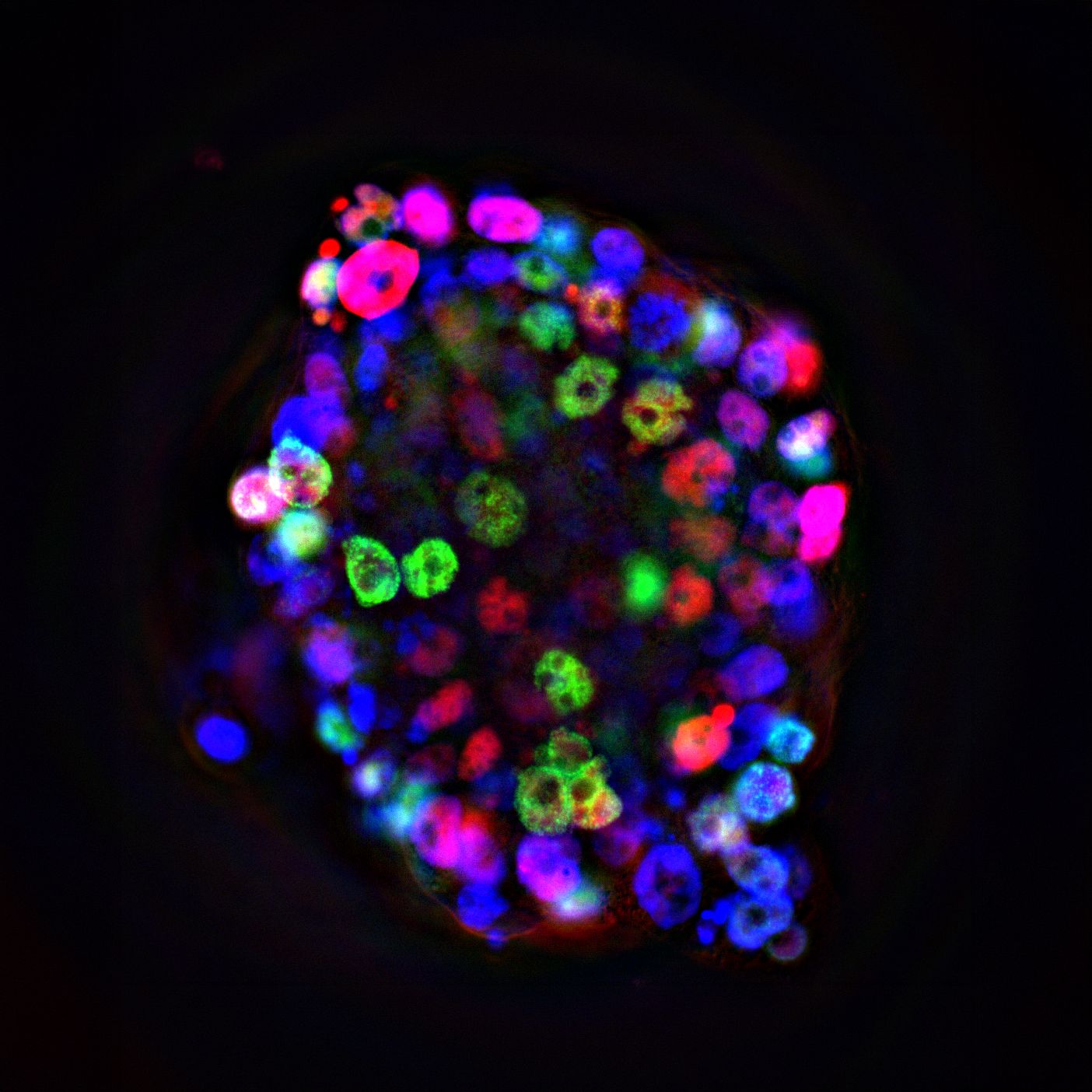
Organoids are 3D structures grown in the laboratory from stem cells or patient-derived tissue that reproduce some of the key features of organs or tumours. Because they retain many of the structural, genetic and biological characteristics of the tissue from which they were derived, organoids can provide a more representative model for studying how cancer develops and responds to treatments.
Building a resource for cancer research
The studies involving Espejo Valle-Inclán and Corbo brought together organoid models from several cancer types, including colorectal, oesophageal, pancreatic, stomach, ovarian, brain, breast and prostate cancers.
Research institutions and hospitals worked together to generate hundreds of models from tumour samples donated by patients. Researchers then compared the organoids with the original tumours, analysing their DNA and patterns of genetic activity to assess how closely the laboratory-grown models reflected the tumours they came from.
The results showed that the models retained many of the important genetic and molecular features of the original tumours, supporting their use as representative models for cancer research.
The researchers then used a range of techniques, including whole-genome sequencing, RNA sequencing and CRISPR screening, to investigate which genes cancer cells depend on to grow and survive. This revealed thousands of genetic dependencies, including vulnerabilities shared across different cancer types as well as others associated with particular tumour types or genetic features.
By combining these results with clinical and genomic information, the researchers could link the molecular characteristics of tumours to the genes and biological processes they depend on. These findings provide new insights into the mechanisms driving different cancers types and may help identify potential targets for future therapies.
The models can also be used to study how cancers evolve over time. In some cases, researchers generated models from the same patient before and after treatment, allowing them to investigate how tumours develop resistance to therapy and to identify vulnerabilities that may emerge as cancer adapts.
Espejo Valle-Inclán and Corbo brought critical computational, organoid generation and biobanking expertise to the HCMI initiative.
Espejo Valle-Inclán played a central role in the bioinformatic pipelines and data analysis for the Sanger Institute organoid biobank and the HCMI flagship studies. His work focused on whole-genome sequencing analysis, essentially mapping the complete DNA instruction manual of the tumours. This involved benchmarking specific spelling mistakes in the DNA, missing or added pieces of genetic code and overall patterns of mutational activity to prove the genomic fidelity of organoids against their patient tumours of origin.
Vincenzo Corbo served, together with Aldo Scarpa, as co-Principal Investigator of the Italian subsite of the NCI-funded Cancer Model Development Center participating in HCMI. Based at the University of Verona’s ARC-Net Research Centre, the team’s role was to establish patient-derived cancer models from surgically resected tumours, with a focus on pancreatic and colorectal cancers. The close integration of surgical pathology, tissue processing and advanced cell culture methodologies allowed the generation of robust, expandable models suitable for molecular characterization, long-term banking and distribution. The Verona team ultimately generated and distributed more than 70 models to the HCMI resource, contributing to the creation of a living collection that could be shared and studied by researchers worldwide.
For Espejo Valle-Inclán, “this resource could help scientists prioritise new therapeutic hypotheses for cancers where better treatments are needed. Rather than replacing traditional 2D cell lines, the organoid biobank complements them, helping researchers address areas where existing models are less informative. By linking patient-derived models with clinical and genomic data, this provides a powerful platform to bridge the gap between discoveries in the laboratory and patients.”
For Corbo, the real achievement of HCMI was not simply generating patient-derived models, but showing that these models can retain key features of the tumours from which they originate while being expanded, quality-controlled and shared with the scientific community. “At Verona, our team contributed more than 70 pancreatic and colorectal cancer models. I still remember the first shipments leaving our laboratory for the HCMI repositories: after years of work behind the scenes, that was when the ambition of the project became tangible. Seeing researchers around the world now using these models to address questions that we could not have anticipated at the outset is the ultimate validation of this effort”, he said.
Applying these approaches to pancreatic cancer
At the Würth Research Centre of Hope for Pancreatic Cancer at the Botton-Champalimaud Pancreatic Cancer Centre, Espejo Valle-Inclán and Corbo are now building on the framework established through these global studies to tackle Pancreatic Ductal Adenocarcinoma, one of the most lethal and treatment-resistant malignancies.
While the Nature publications establish a global benchmark across multiple organ systems, the Würth Research Centre of Hope for Pancreatic Cancer is extending this approach to a deeply annotated living biobank dedicated to pancreatic cancer, with a particular emphasis on advanced diseases that remain comparatively underrepresented in existing model collections. By combining patient-derived 3D organoid derivation, high-depth whole-genome profiling, single-cell analysis and functional screening, the team aims to capture the full spectrum of inter- and intra-patient heterogeneity in pancreatic cancer and to create a resource for investigating tumor evolution, treatment response and therapeutic resistance.
"Although our current work at the Champalimaud Foundation builds upon these published datasets, this international milestone serves as a direct operational blueprint for our future. At the Würth Research Centre of Hope for Pancreatic Cancer, we are establishing a cutting-edge pancreatic cancer organoid platform. By applying these exact multi-omic and functional screening pipelines to our local patient cohort, we hope to translate these global discovery frameworks into direct therapeutic breakthroughs for pancreatic cancer patients", says Espejo Valle-Inclán.
A resource for the research community
The value of the HCMI resource lies not only in the number of models generated, but also in the depth of their characterisation and the wealth of associated genomic and functional data. Making these resources available to the wider research community can help scientists investigate cancer biology, identify potential therapeutic targets and understand why some tumours respond to treatment while others develop resistance.
The data generated through these studies are publicly available through DepMap and CellModelPassports. The organoid models are also being made available to researchers through repositories, namely the Life Science business of Merck KGaA, Darmstadt, Germany and the American Type Culture Collection (ATCC).
Research articles
A tumour-derived organoid biobank maps cancer gene dependencies – including Jose Espejo Valle-Inclán
A compendium of next-generation patient-derived models for diverse cancers – including Vincenzo Corbo and Jose Espejo Valle-Inclán
A dependency map enhanced with next-generation 3D cancer models
Image caption: Pancreatic cancer organoid under the microscope. Blue (DAPI) marks the cell nuclei, green (EdU) highlights actively proliferating cells and red (GATA6) marks cells expressing a pancreatic epithelial lineage marker.
Text by Ana Rita P. Mendes, Communication & Events Manager of the Champalimaud Foundation's Communication, Event & Outreach Team