Noam Shemesh was awarded this grant by the European Research Council to establish cutting edge Magnetic Resonance Imaging methodologies that will provide novel insights onto neural function during health and disease.
Functional Magnetic Resonance Imaging (fMRI) is a powerful tool for studying various neuroscience and biomedical questions. Current fMRI methods work by detecting changes in blood volume and oxygenation level that accompany neural activity. These changes, however, occur over a timescale of seconds, while neural activity occurs within a fraction of a single second. This difference in timescale points out an obvious limitation of current fMRI techniques – they are too slow to resolve many important processes in the brain.
Dr. Noam Shemesh, principal investigator at Champalimaud Centre for the Unknown in Lisbon, has been developing novel techniques that harness the power of MRI to study direct measurements of neural activity. “The difference between classical fMRI techniques and the methods I am working on is two fold. These new methods aim to measure neural activity directly, and not through surrogate processes like blood flow, and as such, they can also potentially detect activation dynamics on a much faster timescale”, says Dr. Shemesh.

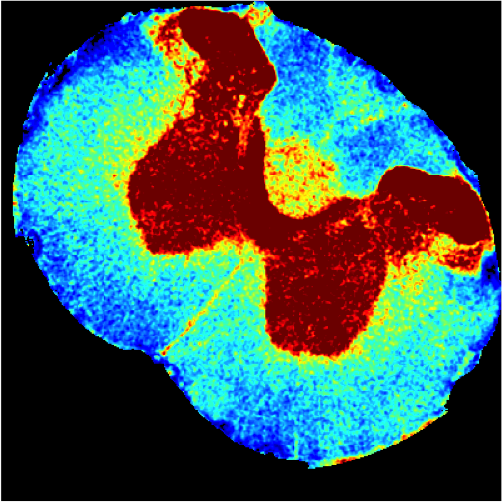
Picture by Francisco Romero.
One of the methods Dr. Shemesh began developing during his postdoctoral work in Israel, measures changes in the volume of neurons. “It is known that when neurons become active, they swell just a little bit. During my postdoctoral studies in Prof. Lucio Frydman’s Lab in the Weizmann Institute of Science, we developed a technique tailored to measuring such subtle changes in neuronal structure. Now, my Lab will develop and apply the technique in fMRI mode, so that these changes can be measured in awake behaving animals.”
Another method Dr. Shemesh developed previously and now intends to implement towards neuroscience research, has the capability of measuring the release of neurotransmitters, another direct measure of neural activity. “The activity of neurons results in the release of neurotransmitters. These small molecules have many functions, including the activation, or the inhibition, of other neurons. Using advanced, ultrahigh magnetic field MRI techniques, I believe we will be able to measure the naturally-occurring dynamics of these neurotransmitters, which will provide us with an unprecedented view onto the inner-workings of neural circuits”, explains Dr. Shemesh.

Dr. Shemesh plans to use these methods to address one of the greatest questions we are currently faced with – how does the brain generate behaviour? “Our ultimate goal is to establish links between behaviour and its underlying global neural circuits.”
These methods will be implemented at the state-of-the-art imaging facilities of Champalimaud Centre for the Unknown. “The Centre has recently acquired cutting-edge ultrahigh field MRI scanners that have established it as one of the most advanced preclinical imaging institutions worldwide. We are looking forward to witnessing unique views into living systems, enabled by unprecedented structural and temporal resolutions the new scanners provide and the novel methodologies we will develop”, concludes Dr. Shemesh.


