06 May 2025
How AI Is Transforming PET/CT Analysis
Medical imaging is at the centre of disease detection, staging and assessment, with clinicians and researchers constantly collaborating to make scans and their evaluation more accurate.
06 May 2025
Medical imaging is at the centre of disease detection, staging and assessment, with clinicians and researchers constantly collaborating to make scans and their evaluation more accurate.

Analysing these scans can also be time-consuming and complex, as doctors need to pore over countless images, looking for often tiny details.
So, any new imaging analysis technique that is faster and more precise is always welcome: a new paper (recently published in the Journal of Nuclear Medicine) reveals that the Champalimaud Foundation’s Nuclear Medicine-Radiopharmacology Unit has managed just that by using Deep Learning (DL) Artificial Intelligence (AI).
The current best-practice in oncology, used at the Champalimaud Foundation, is for a patient to undergo a PET/CT scan, which allows nuclear medicine physicians to observe, in 3-D, the distribution of an injected radiotracer (a radioactive molecule used to track the movement and distribution of a substance within the body). These tomographic images are examined slice by slice in a time-consuming process and then interpreted as a whole, revealing whether, or not, certain tissues may be malignant lesions.
To test how AI could improve scan analysis through automatic and accurate tumour identification, these nuclear medicine researchers applied three different techniques to nearly 1,000 scans.
Focussing on the two most-commonly used radiopharmaceuticals ([18F]FDG - used to detect and monitor cancers like melanoma, lymphoma, and lung cancer; and [68Ga]Ga-PSMA - used to detect and monitor prostate cancer), the researchers utilised AI-based methods to find out if there was a way to streamline and improve the process.
The researchers used a standard deep-learning AI technique for model creation, an enhanced AI model using Maximum-Intensity Projection (MIP) images, and finally a combination of both techniques, to automatically highlight potential tumour sites. Cláudia Constantino, Biomedical Engineer of Nuclear Medicine-Radiopharmacology at the Champalimaud Foundation and first author of this paper identified that the main goal was to: “Automatically identify and outline lesions, with the minimum errors possible, in these essential PET/CT images. This helps nuclear medicine physicians and oncologists to quickly assess tumour size, shape, and spread, making diagnosis and treatment planning faster and more consistent."
For the first step, using a standard deep-learning technique, AI was used to analyse the PET/CT scans directly.
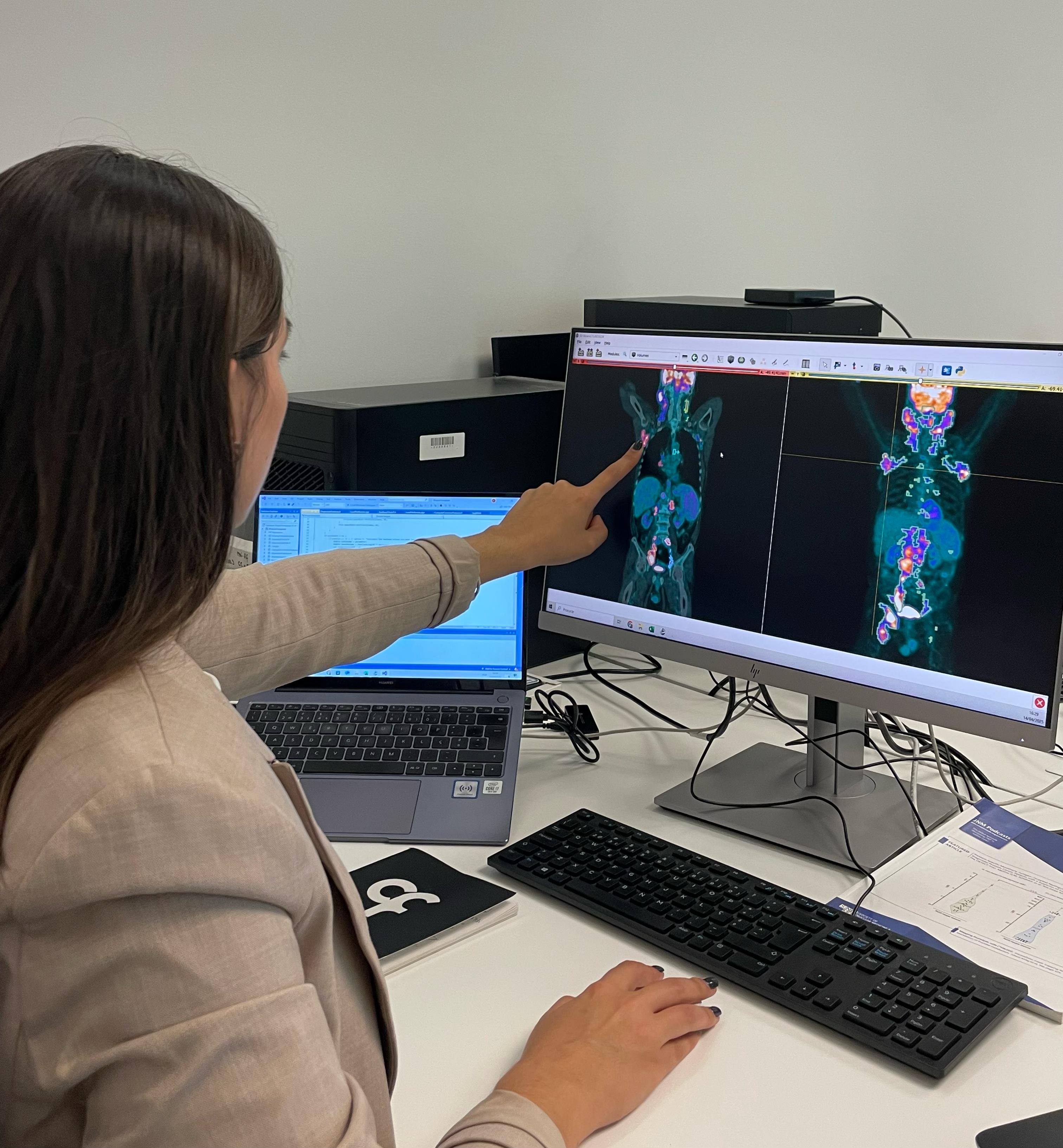
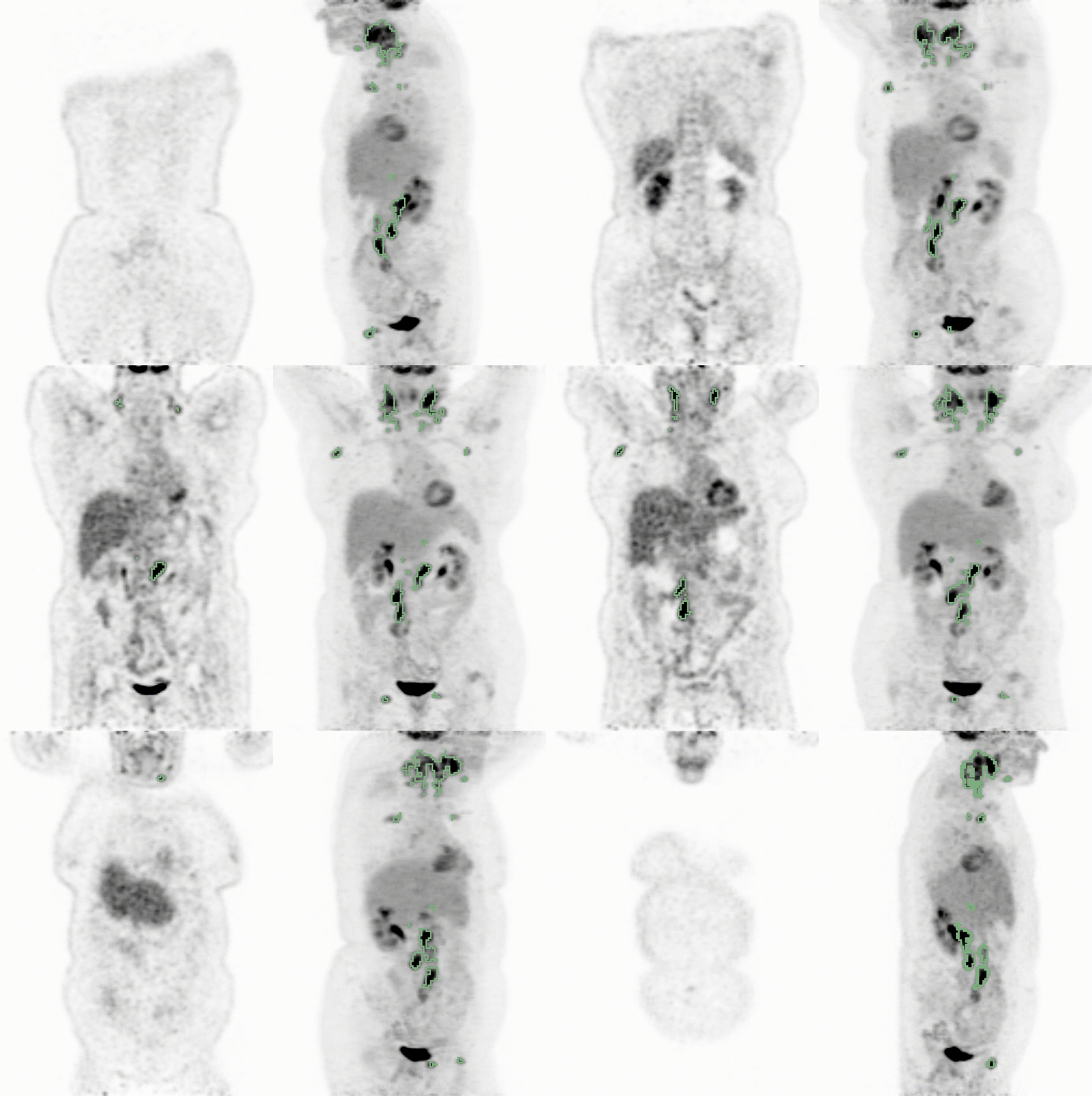
In a second step, from the same PET/CT scans, MIP images were then created. These MIP images use a post-processing technique to condense 3D scan data into a new image where the most intense signals - often tumours - are highlighted, making abnormalities easier to detect. This can be seen clearly in the GIF below: the PET/CT image is on the left, with the corresponding MIP images on the right - both images show the AI-highlighted tumours in green. These MIP images could then be used together with AI to improve the automatic detection of tumours in PET/CT scans.

Finally, a combination of both techniques was used, giving the most compelling results overall in the PET studies. The combination of AI standard model and AI-based MIP imaging model provided the most complete and reliable analysis of the available scans, offering detailed segmentation with almost no false positives - something too time-consuming to achieve in routine clinical practice.
AI-based methods have the potential to be valuable tools for clinicians, helping them analyse scans more quickly and efficiently, and enabling the extraction of more information from the scans. The improved accuracy in measuring tumour burden can provide clearer insights into how a disease is progressing and how well a patient is responding to treatment. Ultimately, AI can serve as an important assistant in medical imaging, helping doctors make faster and more precise decisions while ensuring that patient care remains the top priority.
While AI tools like these can assist nuclear medicine physicians, Cláudia Constantino is very clear on how AI can never replace medical expertise: “Our results showed that we may assist nuclear medicine physicians by taking an initial look at PET/CT images with the help of AI, enabling a complete tumour burden analysis. However, physician supervision remains essential - AI-generated outputs must always be reviewed, corrected, and validated by specialists. Nuclear medicine physicians and oncologists are still essential for interpreting results, making final diagnoses, and considering the patient’s full medical picture.”
For the patient, they can rest assured that their scans contain all of the information that the doctor needs to see, meaning that the medical experts can spend more of their time doing what they do best - diagnosing, treating and caring for their patients.
