Imaging prior to surgery may reduce the risk of incontinence after prostate cancer removal
The removal of localised prostate cancers, which can be life-saving, may come at a price: urinary incontinence. This situation could now be posed to improve, according to recent research.
Radical surgical removal of the prostate is the most frequent treatment for prostate cancer when the tumour is localised (meaning it has not spread outside the prostate).
Nowadays, surgery can be performed by robot-assisted laparoscopy, a minimally invasive and accurate technique. However, urinary incontinence remains one of the main potentially unavoidable complications of the surgery, being responsible for damage to the nerves and muscles of the bladder, urethra and urethral sphincters in 6% to 8% of patients submitted to the procedure. This is due to the fact that the prostate is located just below the bladder and surrounds the urethra, making it difficult to visually distinguish, at the microscopic level, the urinary from the prostatic tissue in order to spare the first during the ablation of the second.
Urinary incontinence has a serious impact on quality of life. And when it does not resolve by itself within a year after surgery, other treatments may be necessary – including a second surgery to insert an artificial urethral sphincter.
Now, a team from the Champalimaud Foundation, in Lisbon, has shown that an advanced magnetic resonance imaging technique, called Diffusion Tensor Imaging (DTI) – could successfully be used to predict, before performing the surgery, the likelihood that an individual patient with prostate cancer will suffer from post-operative long-term urinary incontinence. Their results were recently published in the journal Insights into Imaging.
“Although novel robot-assisted prostatectomy procedures reduce the chance of post-surgical complications,” says Dr. Nickolas Papanikolaou – Head of the Computational Clinical Imaging Group at Champalimaud Research, who led the study – “during prostatectomy the fibers of the proximal and distal sphincters are still damaged in a significant percentage of patients. Studying these structures pre-operatively can provide a quantitative method to predict the rate of post-surgical continence recovery”.
“To our knowledge,” adds Dr. Papanikolaou, “our study is the first to prove the feasibility of DTI, prior to robot-assisted laparoscopy, to visualise, map and measure the properties of the microstructures of each patient’s urethral sphincter complex in a population of prostate cancer patients who are candidates to surgery”.
Indeed, DTI (which has been mostly used to image the brain’s nervous connections) had already proven to allow the imaging of the urethral sphincter complex in healthy young male subjects. But contrary to the subjects involved in those studies, 99% of prostate cancer patients are over 50 years old. This means that their organ tissues are more likely to have suffered the effects of aging, as well as morphologic changes due to the tumour – and this poses further imaging challenges.
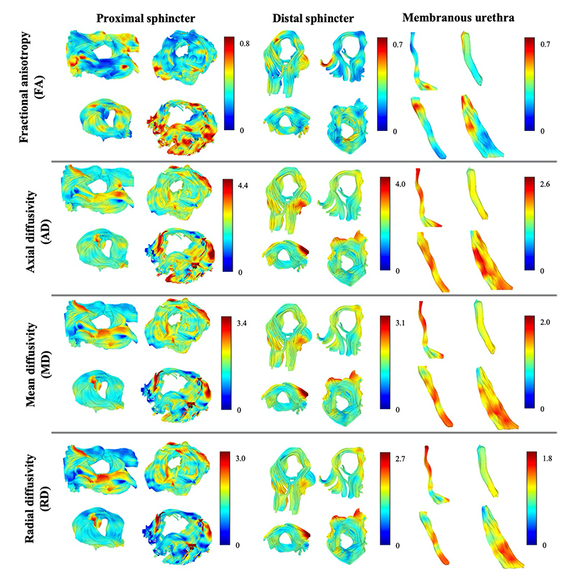
Using DTI, Ana Verde (from Dr. Papanikolaou’s group) and her colleagues reconstructed images of the fibers of the male urethral sphincter complex in 28 prostate cancer patients (with a median age of 64.5 years old) who were set to undergo the laparoscopic procedure at the Champalimaud Clinical Centre’s Urology Unit. More specifically, they showed that the imaging technique, combined with a fiber-tracking algorithm, enabled them to discriminate the urethral sphincters and the urethra from the prostate.
“We have proved that it is possible to use DTI, a non-invasive and in vivo technique, to visualise and quantify the properties of a male individual's urethral complex, in terms of fiber integrity, directionality and density, prior to surgery” explains Dr. Papanikolaou. “Moreover, adding the acquisition of the necessary sequence of DTI images to a standard MRI image acquisition session represents an increment in scanning time of no more than five minutes, while achieving a good image resolution of the fibers.”
“Future steps will be to study whether age influences the DTI measures of the sphincters, to correlate these measures [such as fiber integrity, directionality and density] with post-surgical test scores for incontinence and to test the reproducibility of this technique. We would then use these DTI measures to build a model that could predict the rate of post-surgical continence recovery,” concludes Dr. Papanikolaou.
Ultimately, this may guide patients and clinicians in choosing the best-individualised treatment option – including non-surgical techniques – in order to ensure both the accuracy of tumour removal and the quality of life preservation.
By Ana Gerschenfeld, Health & Science Writer of the Champalimaud Foundation.
Three-dimensional representation of the proximal, distal sphincters and membranous urethra (segment of the urethra external to the prostate), color-coded with information related to biomechanics properties of the muscle fibers, extracted from diffusion tensor imaging data.