When digital surgery meets radiology: Digital Surgery Lab at the European Congress of Radiology 2026
From 4 to 8 March 2026, the global radiology community gathered in Vienna for the European Congress of Radiology 2026 (ECR), one of Europe’s largest medical meetings. This year’s edition, under the theme “Rays of Knowledge”, focused on education, innovation and the continuous exchange of knowledge in the field of radiology.
Among the institutions presenting their work was the Champalimaud Foundation, represented by its Digital Surgery Lab. The multidisciplinary research group, co-led by Tiago Marques and João Santinha, showcased projects at the intersection of medicine, engineering and artificial intelligence (AI).
The lab brings together surgeons, radiologists, engineers and researchers who collaborate to design and test digital solutions, from AI models to extended reality platforms, aimed at transforming how surgery is planned and performed. Their presence at the ECR 2026 highlighted a broader shift in medicine: the growing integration of imaging, digital technologies and surgical decision-making.
Across conference sessions and a live demonstration, the team presented new approaches to AI-driven healthcare, tumour visualisation in breast cancer surgery and digital tools that can reshape clinical workflows. The discussions reflected a shared ambition within the field: to move beyond technological innovation and toward solutions that can be implemented at scale in everyday clinical practice.
2050: Are you ready for AI-driven healthcare?
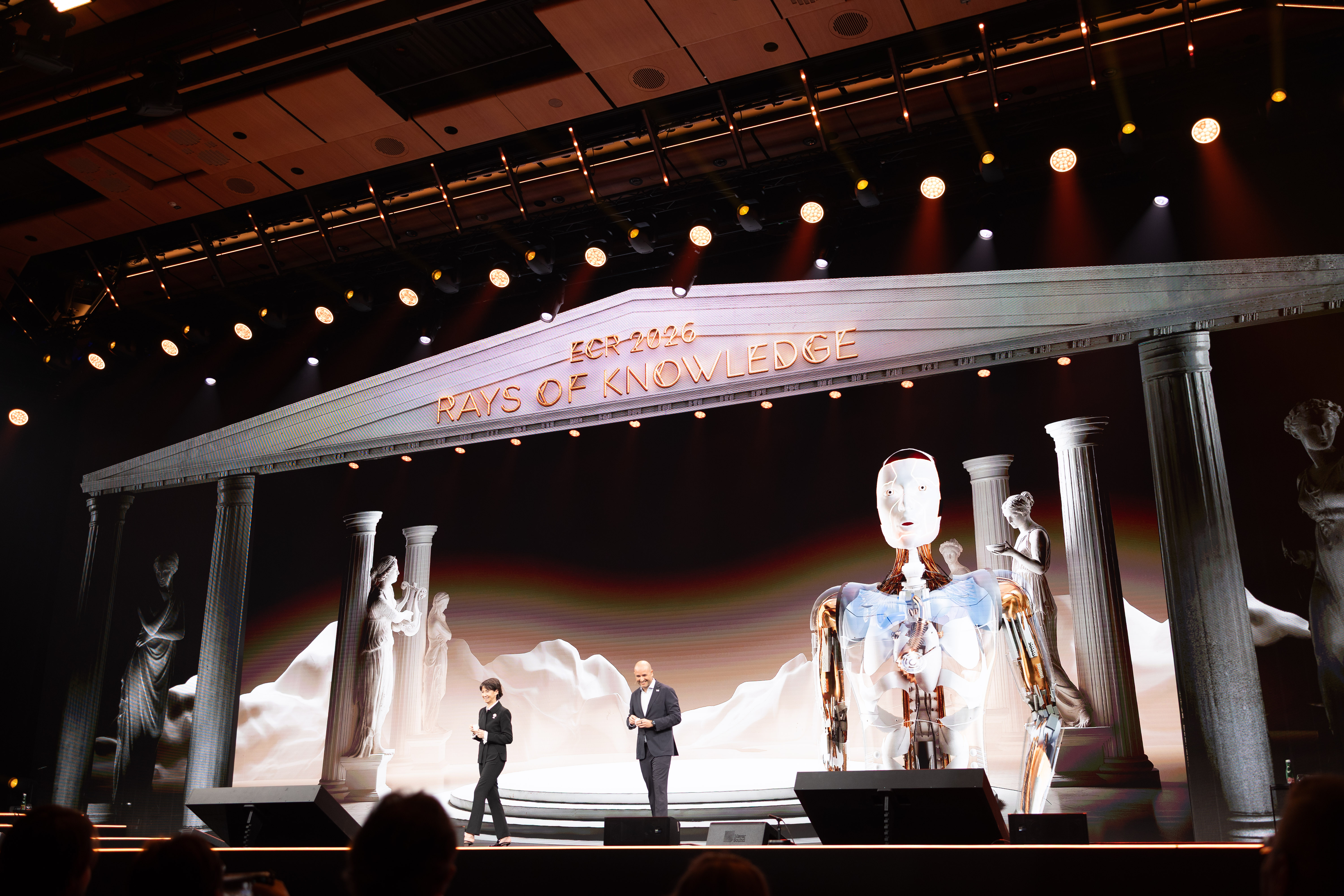
One of the most anticipated contributions from the group took place during a joint session entitled “2050: Are you ready for AI-driven healthcare?” The talk, delivered by surgeon-researcher Pedro Gouveia, GSL chief-medical surgeon, alongside radiologist Regina Beets‑Tan, Group Leader at the Netherlands Cancer Institute, explored how AI can reshape the healthcare landscape in the coming decades.
Although AI has become a dominant theme in medical research and innovation, Pedro Gouveia emphasised that the real challenge lies not in the technology itself but its adoption. “There is still a large gap between AI-based technological innovation and its implementation,” he explained. “It’s very common to ask someone what AI has done for them and the answer is ‘nothing’, and they’re probably right.”
Barriers such as limited infrastructure and low technological literacy continue to slow the translation of AI tools into everyday clinical practice. For the panelists, preparing healthcare professionals, institutions and patients for this transition is essential. Pedro often compares the challenge to the early days of mobile communications. “What we want from the implementation of AI in healthcare is the same thing that happened with mobile phones in the 1980s – the democratisation of access regardless of socioeconomic class,” he said. “That’s what scalable implementation means.”
The idea for the joint session emerged from a shared desire to address both the technological and cultural dimensions of AI adoption. Rather than focusing on algorithms or datasets, the talk explored how clinicians and health systems must adapt to work alongside intelligent tools. “Normally humans fear what they don’t know,” Pedro noted. “We wanted to present a new perspective on AI that doesn’t scare people but helps them see it as something that enhances our capacity to prevent, diagnose and treat patients.”
To illustrate this partnership between humans and machines, the speakers introduced a digital collaborator named Ave, as a symbolic member of their team. Together, the trio – surgeon, radiologist and AI agent – demonstrated a future in which clinical expertise and digital intelligence work side by side.
Regina Beets-Tan summarised the potential impact of AI on clinical practice in simple terms: “AI will help us to be more efficient. It will help with standardising, but also with becoming more accurate in our diagnosis and in our workflow with clinicians. It will help us predict the response to treatment.”
For both speakers, the future of medicine will not replace human expertise – it will amplify it.
Joint session "2050: Are you ready for AI-driven healthcare?" – Regina Beets-Tan and Pedro Gouveia
Predicting response and recurrence in breast cancer
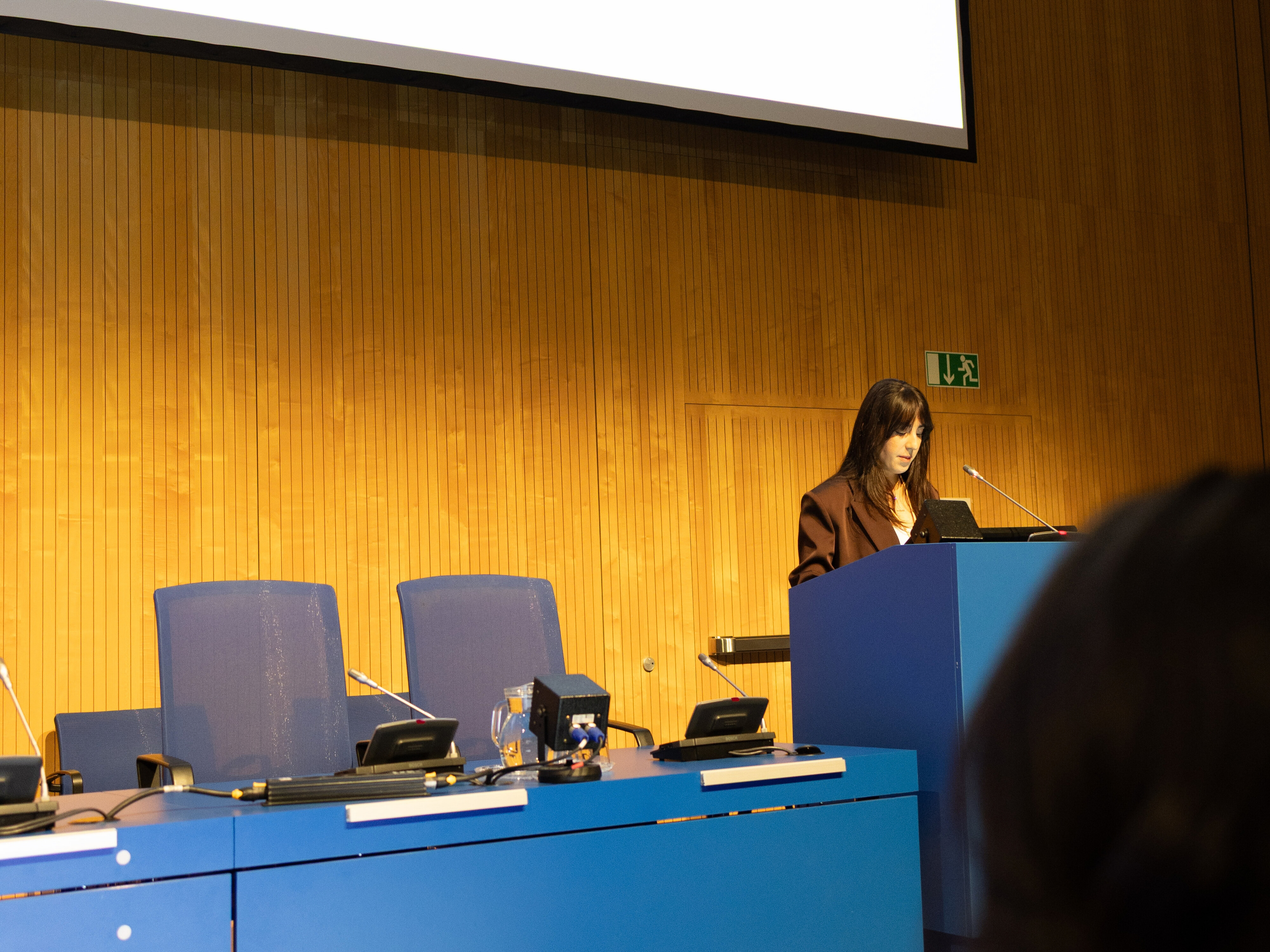
Another highlight of the lab’s participation was the presentation by PhD candidate Rafaela Timóteo. Her research addresses a longstanding clinical challenge: accurately locating tumours during surgery when the lesions are small and often non-palpable.
Current localisation techniques rely on invasive methods such as wires or markers inserted into the tumour before surgery. These approaches can be uncomfortable for patients and do not always provide precise information about tumour boundaries.
Rafaela’s work explores an alternative approach based on extended reality technologies combined with patient-specific digital models. “The research I presented at the ECR reflects the work our lab has developed over the last few years,” she said. “It focuses on improving preoperative and intraoperative planning in breast-conserving surgery, particularly tumour localisation.”
The process begins with two magnetic resonance imaging (MRI) scans, one acquired while the patient lies prone and another while lying supine. Using these images, Rafaela and her colleagues generate a digital twin of the patient’s breast, including fat tissue, fibroglandular structures and the tumour itself.
This digital model is then visualised through extended-reality technology and automatically aligned with the patient in the operating room using depth sensors. The result is a three-dimensional visualisation of the tumour projected onto the patient’s anatomy, enabling surgeons to see exactly where the lesion lies.
Traditional localisation techniques provide guidance but often require surgeons to mentally reconstruct a three-dimensional anatomy from two-dimensional images. The new approach aims to reduce that cognitive burden. “By integrating detailed anatomical information directly into the surgical field, we can enhance spatial understanding,” she explained.
Early results suggest that combining extended reality with digital twins is technically feasible and can enable accurate, non-invasive tumour localisation. The ultimate objective is to help surgeons achieve clear surgical margins, reducing the need for additional operations. “By lowering the cognitive load for surgeons, this approach allows them to make better-informed decisions with greater precision,” Rafaela said.
Before the technology can reach the clinical practice, further validation is required. Future work will involve larger patient cohorts and additional technological refinements, including improved biomechanical models that account for tissue deformation during surgery and automated image segmentation powered by machine learning. “The opportunity to present this research at the ECR was a major milestone in my PhD,” she said. “This work reflects the collective effort of our lab over several years and sharing it at such a respected conference was both rewarding and meaningful.”
Research talk "Predicting response and recurrence in breast cancer" – Rafaela Timóteo
The Cube: demonstrating digital surgery
Beyond conference presentations, the Digital Surgery Lab also participated as an exhibitor, in the Cube – an interactive environment dedicated to hands-on learning in interventional radiology – showcasing their setup to improve preoperative and intraoperative planning in breast-conserving surgery, particularly tumour localisation. Their demonstration gave attendees the opportunity to interact directly with their emerging technology, marking the first time Portugal had representation in the exhibitors area of the ERC.
The demonstration was led by researchers Tiago Marques, João Santinha, Nuno Loução and Rafaela Timóteo, and surgeon Pedro Gouveia. “With this 3D information from the MRI scans, we can superimpose the tumour onto the patient’s body in the operating room,” João noted. “This allows surgeons to see exactly where the tumour is and what the margins are.” The potential benefit is significant: more accurate tumour removal and fewer repeat surgeries.
Although the technology was developed primarily for surgical applications in patients with breast cancer, the radiology audience at the ECR quickly identified additional possibilities. Several participants suggested that similar tools could support image-guided biopsies or other interventional procedures. For example, in liver biopsies, clinicians often rely on multiple Computed Tomography (CT) scans to confirm the needle positioning. A real-time visualisation system could simplify this process while reducing radiation exposure and improving the patient experience.
For João Santinha, this first demonstration at the congress exceeded expectations. With over 80 attendees visits per day, many participants returned with colleagues to try the system themselves, often expressing curiosity and enthusiasm about its potential applications.
Demonstration of the Digital Surgery Lab at the Cube – Tiago Marques, Nuala Healy and Rafaela Timóteo
An atmosphere of collaboration
Throughout the week, researchers interacted with clinicians, engineers and industry representatives interested in digital surgery and medical imaging technologies. These conversations ranged from technical discussions about AI integration to broader reflections on how emerging tools might reshape healthcare systems, serving as catalysts for collaboration.
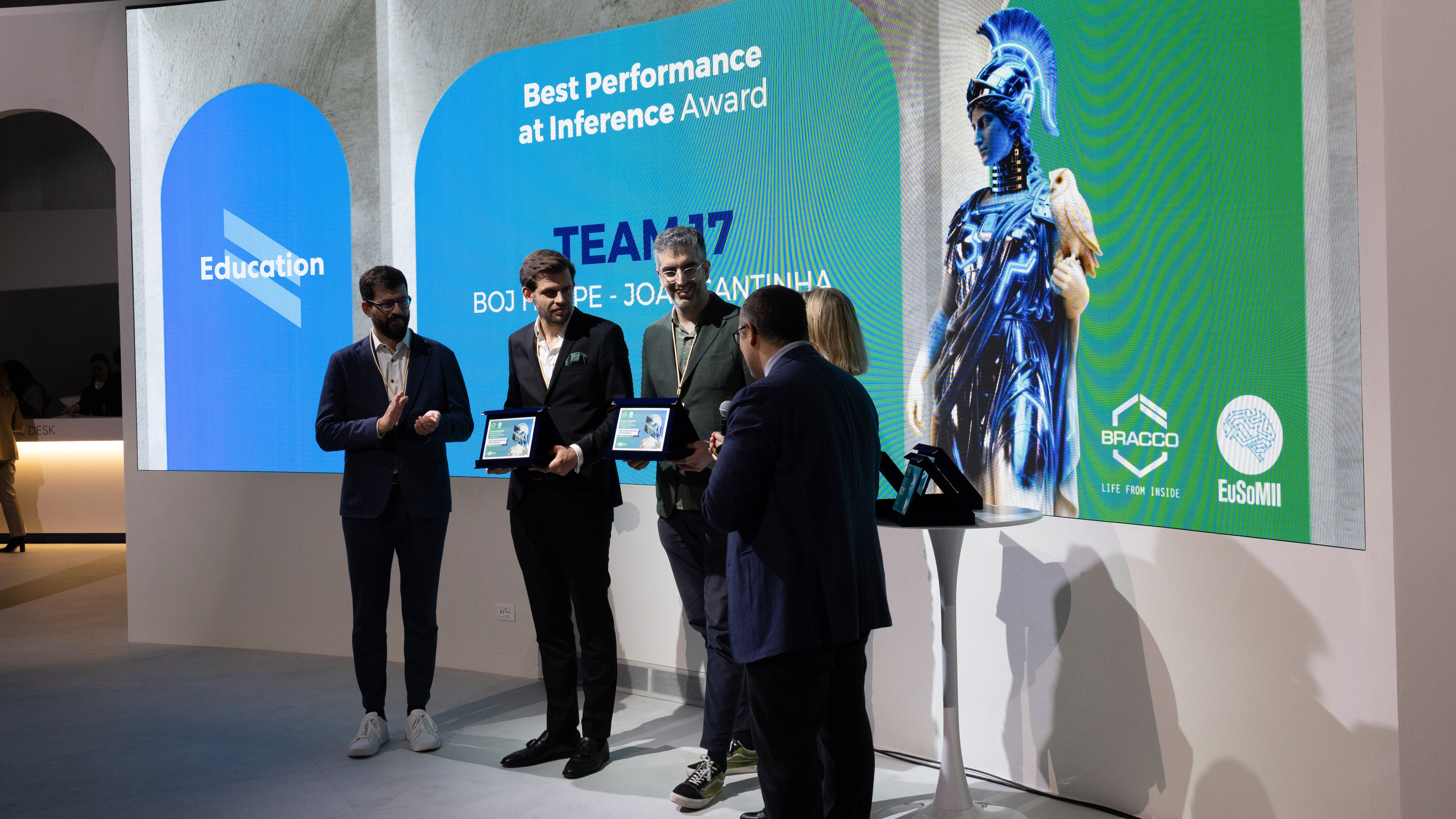
One of the highlights of the week was the participation of João Santinha on the “Hands-on challenge in Agentic AI” hosted by Bracco and EuSoMII at the ECR. This workshop was an important driver of education and professional growth among technicians, radiologists and data scientists by providing them with the opportunity to work together. João Santinha won two awards, one for the “Best performance at Inference” and another for “Best use of budget”.
For the lab members present, this experience was particularly valuable as their work depends on cross-disciplinary expertise. Integrating imaging data, computational modeling and clinical workflows requires collaboration between specialists who traditionally operate in separate domains. In this sense, the ECR served not only as a platform for showcasing innovation but also as a meeting point where future research directions can began to take shape.
Bracco and EuSoMII award for the best performance at inference – João Santinha
Looking ahead
The conversation around AI and digital medicine will continue at the next edition of Medica AI, which will take place at the Champalimaud Foundation on 16 July 2026. The event will bring together experts exploring the future of AI and innovation in medicine.
Information about the speakers and tickets can be found here.
To learn more about the group and its ongoing projects, visit on the Digital Surgery Lab page.
Text by Diana Cadete, Events & Engagement Manager of the Champalimaud Foundation's Communication, Events & Outreach Team